
Page 69 - OxyBand Research Background
P. 69
T
ASE REPOR
ORIGINAL RESEARCH
C
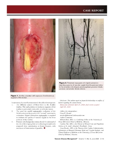
Figure 2. Peripheral angiogram with digital subtraction
depicting a paucity of vascular supply from the peroneal artery
to the ulceration site despite patent popliteal, posterior tibialis,
peroneal, and anterior tibialis arteries.
Figure 1. Achilles ulceration with exposure of mid-tendinous
segment of the tendon.
Disclosure: The authors report no financial relationships or conflicts of
In summary, this case illustrates several clinically relevant points: interest regarding the content herein.
1. The different sources of blood flow to the Achilles Manuscript submitted April 25, 2020, final version accepted
tendon. The mid-portion or tendinous segment of the April 28, 2020.
tendon is particularly vulnerable to ischemic injury.
2. The need for careful angiographic evaluation of the Address for correspondence:
Achilles ulcers in the presence of a “normal” noninvasive Oscar R. Rosales
evaluation. Digital subtraction angiography is required orosales@HoustonCardiovascular.com
to evaluate the quality of vascular supply to the three Author Comments:
portions of the tendon. Michael Hust, MD, is a Cardiology Fellow at the University of
3. Patients with benign skin lesions should be informed of Texas-McGovern School of Medicine, Houston.
the risk of developing slow or nonhealing wounds with Caroline Fife, MD, is the Director of Wound Care and Hyperbaric
proposed deep cauterization or frozen biopsies, with Services at St. Luke’s Hospital, Woodlands, Texas.
avoidance of intervention if possible. n Oscar Rosales, MD, is the Director of the Cardiac Catheterization
Laboratories at Memorial Hermann Heart and Vascular Institute, and
Clinical Professor of Medicine at the University of Texas-McGovern
School of Medicine, Houston.
Vascular Disease Management Volume 17, No. 6, June 2020 E118
®